FURTHER EVALUATION OF LOWER BACK PAIN SYMPTOMS
Advanced imaging and related tests
CAT Scan or CT
A CT scan of the low back is a special X-ray image that takes 360 degree pictures of the spine. A CT of the lumber spine offers much more detail that the standard X-ray picture discussed above. However, CT scans also suffer some of the same limitations as X-rays.
MRI
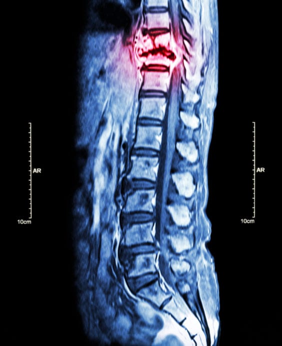
An MRI provides detailed images of nerves, muscles, and tendons by using magnetic fields and radio waves. Unlike the limitations seen with X-rays, MRI images are able to identify specific “pinched nerves”. A major additional advantage is that MRI results in no radiation exposure to patients. A major disadvantage is that MRI may not be able to be used in those with pacemakers, defibrillators, or other metal implants.
Myelogram
A special contrast dye is injected into the spine and CT scans are performed following the injection. A detailed image of the spinal column is created for review by lower back pain doctors. This imaging technique requires that a specially trained physician be available to directly inject the contrast dye into the spine. Most routine imaging facilities may not have access to this specific type of spine testing.
Discogram
A discogram can determine which specific disc is the cause of a patient’s lumbar pain. This technique is used when surgery is being considered. The overwhelming majority patients with back pain will not require or benefit from this particular study. It is used for specific types of back pain conditions.
EMG and/or NCV
Electromyography (EMG) measures a muscles response to a small electrical stimulation. This test helps identify which muscles and nerves are being impacted by a problem within the spine It is performed by a physician specialist to find which nerves of the lumbar spine are “pinched”.

 CALL TODAY!


